
3D Cone Beam CT Imaging
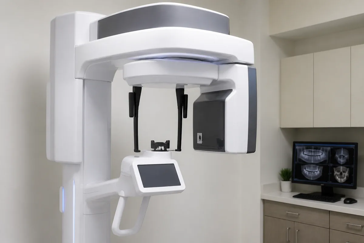
3D Cone Beam CT Imaging
3D Cone Beam CT (CBCT) imaging is a specialized type of dental X-ray that captures a three-dimensional view of your teeth, jawbone, nerve pathways, and surrounding soft tissue in a single scan. At 1st Family Dental in Chicago, IL, this imaging is used to support precise, well-planned treatment for implants, oral surgery, and other cases where a flat, two-dimensional X-ray doesn't tell the whole story.
What 3D Cone Beam CT Imaging is
Traditional dental X-rays produce a flat, two-dimensional picture. That works well for spotting cavities or checking a single tooth, but it can't show depth, the exact position of a nerve, or how thick and dense the bone is in a particular spot. Cone Beam CT imaging solves this by rotating a cone-shaped X-ray beam around the head and capturing hundreds of images, which are then reconstructed into a detailed 3D model.
The result is a single scan that shows teeth, soft tissue, nerve paths, and bone density together, giving a much fuller picture than a series of standard X-rays. Because the scan is digital, it can be viewed, rotated, and measured on a screen, which makes it easier to plan surgical or implant procedures with more certainty before any work begins.
How we use it at 1st Family Dental
At 1st Family Dental, 3D Cone Beam CT imaging is used mainly to plan treatment that involves the jawbone or its surrounding structures — most often dental implants, All-on-4 implants, bone grafting, sinus lifts, wisdom teeth removal, and other oral surgery. Several members of our clinical team have training that specifically involves 3D imaging and CT-guided planning. Dr. John Hur, an Oral and Maxillofacial Surgeon, has training that includes CT-guided skeletal mapping and surgical protocols for implant and corrective jaw surgery. Dr. Najeeb Abufarha's training includes 3D imaging as part of his work in implant placement, guided surgery, and bone grafting. Among our orthodontists, Dr. Sameer Al-Obaidi and Dr. Budi Kusnoto both have training and experience using 3D imaging in diagnosis and treatment planning, including for cases involving temporary anchorage devices or surgical orthodontics.
CBCT scans are often used alongside other on-site tools. For implant cases, the scan may be paired with our Guided Implant Surgery process, where the treatment plan built from the 3D image guides the actual placement in the mouth. An iTero Element digital scan of the teeth and gums, or a Digital Smile Preview, may also be used together with CBCT data to plan how a final result will look and function.
What it means for you
A 3D scan gives your dentist or surgeon a clearer, more complete picture before any surgical work is planned, which supports more accurate treatment planning. It can also mean fewer separate X-rays are needed, since one CBCT scan captures information that might otherwise take several images to piece together.
Because the scan is quick and non-invasive, it's generally comfortable for patients, and reviewing the 3D model together with your dentist can make it easier to understand your own anatomy and the reasoning behind a recommended treatment plan.
What to expect during your visit
No special preparation is usually needed before a Cone Beam CT scan. You'll be asked to remove any metal objects near your head or neck, such as glasses, earrings, or a removable dental appliance, since metal can interfere with the image. You'll then stand or sit in the scanner while it rotates around your head; you'll need to stay still, but the scan itself typically takes well under a minute.
Afterward, the images are processed into a 3D model that your dentist or specialist can review with you on a screen, pointing out the specific areas relevant to your treatment — such as bone density where an implant is planned, or the position of a nerve near a wisdom tooth. From there, the scan becomes part of your treatment plan and may be referenced again at later visits as care progresses.
Is 3D Cone Beam CT Imaging safe?
Cone Beam CT imaging uses X-rays, similar to standard dental imaging, and is a digital technology designed to capture detailed information efficiently. As with any X-ray, your dental team will weigh the benefit of the additional detail against the reason for the scan, and will explain why a CBCT scan is being recommended for your specific situation.
How long does the scan take?
The scan itself is fast, generally completed in under a minute while you remain still in the machine. The full appointment may take a bit longer once you account for positioning and reviewing the images afterward, but the imaging portion is brief compared to the planning conversation that follows.
Who typically needs this type of imaging?
CBCT imaging is most often used for patients being evaluated for dental implants, bone grafting, sinus lifts, wisdom teeth removal, or other oral surgery, as well as some orthodontic cases involving surgical planning or temporary anchorage devices. Your dentist will let you know if your treatment calls for this type of scan.
If you'd like to learn more about whether 3D Cone Beam CT imaging is part of your recommended treatment plan, reach out to 1st Family Dental to ask us about it.